RETINAL DETACHMENT
What is the retina?
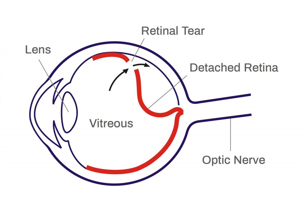
Imagine that your eye is like a camera, and the retina is a photographic film. The retina is a fine sheet of nerve tissue lining the inside of the eye (see diagram). Rays of light enter the eye and are focused onto the retina by the lens. The retina produces a picture, which is sent along the optic nerve for the brain to interpret. It’s rather like the film in the camera being developed so that pictures can be produced.
What is retinal detachment?
Retinal detachments often develop in eyes with retinas weakened by a hole or tear. This allows fluid to seep underneath, weakening the attachment so that the retina becomes detached – rather like wallpaper peeling off a damp wall.

When detached, the retina cannot compose a clear picture from the incoming rays and vision becomes blurred and dim.
What are the symptoms?
The most common symptom is a shadow spreading across the vision of one eye. You may also experience bright flashes of light and/or showers of dark spots called floaters. These symptoms are never painful.
Many people experience flashes or floaters and these are not necessarily a cause for alarm. However, if they are getting severe and seem to be getting worse and you are losing vision, then you should seek medical advice.
Prompt treatment can prevent blindness
Who is at risk of retinal detachment?
The detachment of the retina is more frequent in middle-aged, short-sighted people. However, it is quite uncommon and only about one person in ten thousand is affected. It is rare in young adults.
What is the treatment?
If you get help early, it may only be necessary to have laser or freezing treatment. This is usually performed under local anesthesia. Frequently, however, an operation will be needed to repair a hole or put the retina back in place. In around 80 per cent of cases, the retina can be repaired with a single operation. One in five patients requires more than one surgery to fix the retina. The operation does not usually cause much pain, but your eye will be sore and swollen for
a few days afterwards. Typically, you will be in the hospital for a few hours on the day of the operation. We want to reassure you that the surgeon does not take your eye out of its socket to operate on it.
To learn more about vitrectomy procedure for retinal detachment go here
How much vision can I expect after a successful operation?
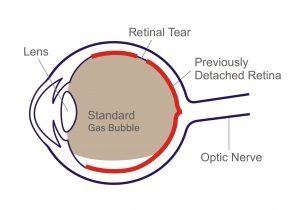
This depends on how much the retina has detached and for how long. The shadow caused by the detachment will usually disappear when the retina has been put back in place. If your ability to see fine detail has been damaged before the operation (ie the macula if off), the central vision may not fully recover after surgery. If a gas bubble is injected in the eye, your vision will be blurred for a couple of weeks.

Gas Bubble after retinal detachment surgery
What happens after the operation?
You will be encouraged to get up and carry on as usual on the day after the operation, although sometimes you will be asked to keep your head in a particular position to help the healing process. Your eye specialist will prescribe eye drops and you will need to use these for a few weeks.
What happens if the detached retina is not put back in place?
Most people will lose all useful vision if no operation is carried out, or if the treatment is unsuccessful. However, further treatment is usually possible if it does not succeed the first time.
Can retinal detachment be prevented?
If your family has a history of retinal detachment, or your doctor finds a weakness in your retina, then preventive laser or freezing treatment may be needed. However, in most cases, it is not possible to take preventive action.
Retinal detachment does not happen as a result of straining your eyes, bending or heavy lifting.
What about my other eye?
If you have had a retinal detachment in one eye, you are at an increased risk of developing one in the other eye. However, there is only about a one in ten chance of this happening. If you have weak areas in your retina, the doctor may advise you treatment to strengthen your retina.


