")
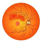
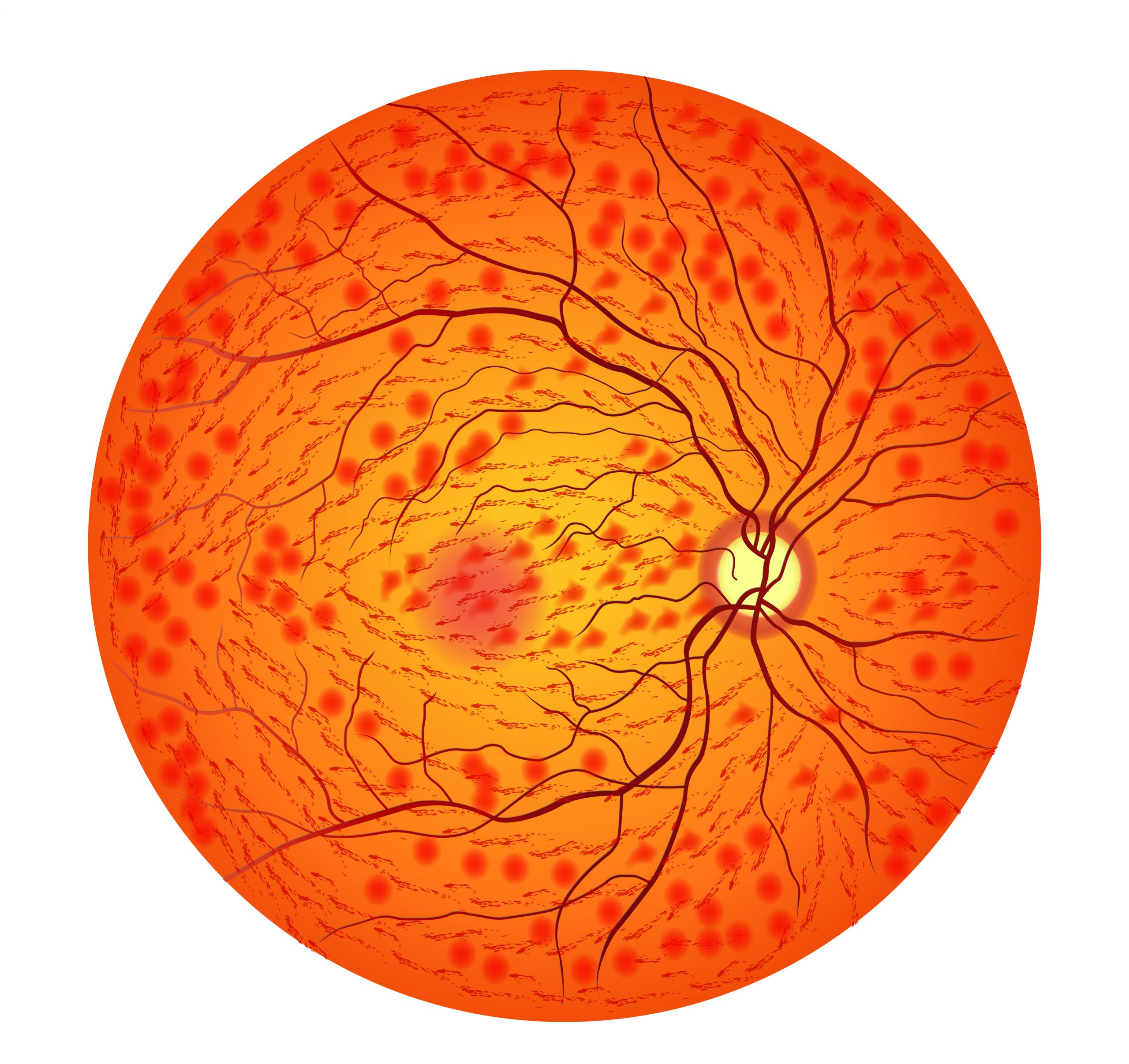
Central Retinal Vein Occlusion (CRVO)
What is a Retinal Vein Occlusion (RVO)?
Retinal vein occlusion (RVO) is a blockage or occlusion of a vein that carries blood away from the retina of the eye. The retina is the tissue located at the back of the eye responsible for vision. When the vein is blocked, blood and fluid accumulate in the retina, leading to reduced vision or even vision loss.
What are the types of RVO?
There are two types of RVO: central retinal vein occlusion (CRVO) and branch retinal vein occlusion (BRVO).
- Central retinal vein occlusion (CRVO) occurs when the main vein that drains the entire retina is blocked.
- Branch retinal vein occlusion (BRVO) occurs when one of the smaller veins branching off the main vein becomes blocked.
What are the symptoms of CRVO?
The symptoms of CRVO can vary depending on the severity and type of the occlusion. However, the common symptoms include:
- Sudden, painless vision loss or blurred vision
- Seeing floaters or dark spots in the vision
- Distorted or wavy vision
- Loss of peripheral vision
- Eye pain
How prevalent is CRVO, and who is at risk?
CRVO is a relatively uncommon condition, affecting around 1% of the general population. The risk of developing CRVO increases with age and in people with certain underlying medical conditions, including:
- High blood pressure
- Diabetes
- Glaucoma
- Atherosclerosis (narrowing of blood vessels)
- Blood clotting disorders
- Elevated cholesterol levels
- Cardiovascular disease
Other risk factors for CRVO include smoking, obesity, and a family history of the condition.
How is CRVO diagnosed?
CRVO is diagnosed through a comprehensive eye exam by an ophthalmologist or optometrist. The exam may include:
- Visual acuity test: This is a standard eye chart test to measure vision.
- Dilated eye exam: The doctor will use drops to dilate the pupil and examine the retina.
- OCT Test: This tests evaluates retinal swelling (macular edema) caused by vein occlusion. If macular edema is established on OCT test, it is an indication for treatment.
- Fluorescein angiography: This is a specialized test where a dye is injected into the arm, and photographs are taken of the retina to assess blood flow and detect blockages.
What are the treatment options for CRVO?
The treatment for CRVO depends on the severity and type of the occlusion. The aim of treatment is to reduce swelling and prevent further vision loss.
- Observation: In some cases -when there is no retinal swelling- the doctor may recommend close monitoring of the condition to see if it resolves on its own.
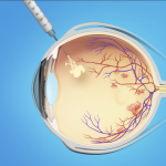
- Injections: Most patients with reduced vision and macular edema will need anti-VEGF injections into the eye to reduce swelling and improve blood flow.
- Medications: Medications may be prescribed to reduce swelling, including anti-inflammatory drugs, steroids, and blood thinners.
- Laser therapy: Laser therapy is a procedure where a high-intensity beam of light is used to seal leaky blood vessels and reduce swelling.
Most patients with reduced vision and macular edema will need multiple anti-VEGF injections into the eye to reduce swelling and improve vision.
Are the procedures painful?
Some treatments for CRVO, such as laser therapy and injections, may cause some discomfort. However, the doctor will usually use a local anesthetic to numb the area before the procedure to minimize any pain or discomfort.
What are the complications of CRVO treatment?
Some of the potential complications of CRVO treatment include:
- Infection
- Bleeding
- Increased eye pressure
- Cataracts
- Retinal detachment
Your doctor will discuss the potential risks and benefits of any treatment options with you before proceeding with the treatment.
What happens if CRVO is left untreated?
If CRVO is left untreated, the blockage can lead to permanent vision loss. In severe cases, the eye pressure may go up causing a painful blind eye.
Untreated CRVO may lead to a blind painful eye.