
Macular Hole
What is a macular hole?
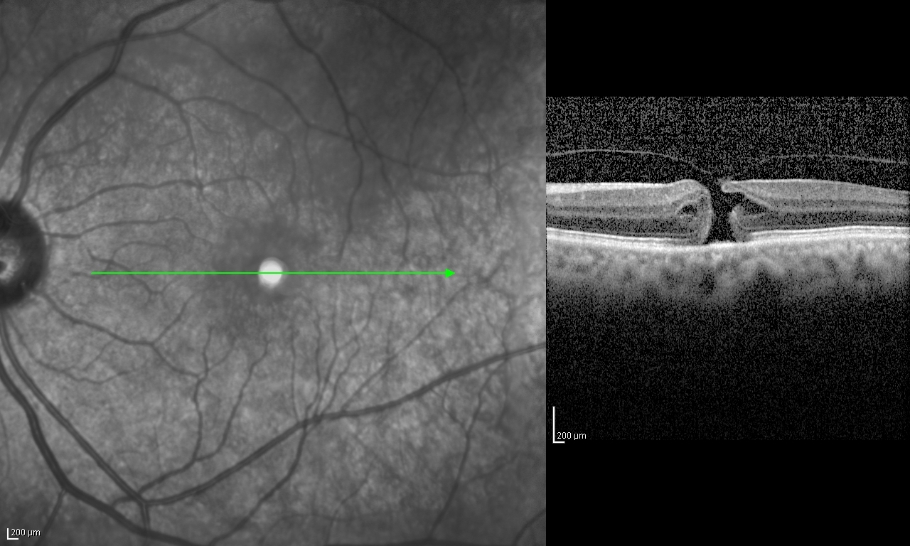
Macular hole is a full thickness gap in the centre of the most central part of the eye called fovea. It usually presents with reduced vision or central distortion. Risk factors include old age, trauma, use of Tamoxifen and female gender. There are 4 stages of macular hole. The most useful test to detect macular hole is OCT. The above OCT scan shows a stage II full-thickness macular hole.
What are the symptoms of a macular hole?
The symptoms of a macular hole can vary from person to person, but some common symptoms include:
- Blurred or distorted vision in the central part of your visual field
- Difficulty seeing fine details, such as reading small print or recognizing faces
- A dark or blank spot in the center of your vision
- Straight lines appearing bent or wavy
If you experience any of these symptoms, it is important to schedule an appointment with a retina specialist as soon as possible.
What causes a macular hole?
In most cases, a macular hole is caused by the natural aging process of the eye. As we age, the vitreous gel that fills the inside of the eye begins to shrink and pull away from the retina. This process is called posterior vitreous detachment (PVD), and it can sometimes cause a small tear to form at the fovea.
Other risk factors for macular holes include:
- Female gender
- Trauma to the eye
- High myopia (nearsightedness)
- Tamoxifen use
How is a macular hole diagnosed?
To diagnose a macular hole, an eye doctor will perform a comprehensive eye exam, which may include the following tests:
- Visual acuity test: This test measures how well you can see at different distances.
- Dilated eye exam: During this exam, your doctor will use special drops to dilate your pupils, allowing them to examine the inside of your eye.
- Optical coherence tomography (OCT): This test uses light waves to create detailed images of the retina, which can help your doctor see if there is a hole in the macula.
How is a macular hole treated?
The treatment for a macular hole depends on the size and severity of the hole. In some cases, a small macular hole may heal on its own without any intervention. However, if the hole is larger or causing significant vision problems, your doctor may recommend one of the following treatments:
- Vitrectomy surgery: This is a surgical procedure in which the vitreous gel inside the eye is removed and replaced with a gas bubble, which helps to close the hole in the macula. The gas bubble stays in the eye for8-9 weeks. This procedure is usually done under local anesthesia as a day care procedure.
- Face-down positioning: After vitrectomy surgery, your doctor may recommend that you maintain a face-down position for a period of time to help the gas bubble stay in place and promote healing of the macular hole. Depending on the size of the macular hole, this may vary from 24 hours to 7 days.
- Observation: In some cases, your doctor may recommend a “watch and wait” approach, monitoring the macular hole over time to see if it gets better or worse.
The anatomical success rate of macular hole surgery is more than 90%. Visual recovery depends on the duration of the hole. Hence, most patients of macular hole are operated fairly quickly, usually within a couple of days. Visual recovery continues for 3-6 months.
How long to I have to keep face down?
Dr Rehman Siddiqui mostly postures his patients face down for 24 hours only. Based on his published research click here cases with small macular hole size <400u do not require long face down posturing to improve macular hole closure.
What do expect after surgery?
- Blurred or distorted vision: It is normal to experience some blurriness or distortion in your vision immediately after surgery. This is usually caused by the gas bubble in your eye, which can cause the image you see to be slightly out of focus. Over time, the gas bubble will dissolve and your vision should gradually improve.
- Eye discomfort: You may experience some discomfort or irritation in your eye after surgery, such as itching or a feeling of pressure. Your doctor may prescribe medication or recommend using cold compresses to help alleviate these symptoms.
- Face-down positioning: If your doctor recommends face-down positioning after surgery, this can be challenging and uncomfortable. You may need to maintain this position for several days or weeks, depending on the size and location of your macular hole. Your doctor will provide specific instructions on how to maintain this position and may recommend certain devices, such as a special chair or mirror, to make it easier.
- Restrictions on activities: After surgery, you will need to avoid certain activities, such as lifting heavy objects or participating in contact sports, to prevent complications and allow your eye to heal properly. Your doctor will provide specific instructions on what activities to avoid and for how long.
- Follow-up appointments: After surgery, you will need to attend follow-up appointments with your doctor to monitor your progress and ensure that your eye is healing properly. Your doctor may perform additional tests, such as OCT or visual acuity tests, to assess your vision and determine if any further treatment is needed.
It is important to follow all of your doctor’s instructions carefully and attend all of your scheduled appointments to ensure the best possible outcome after macular hole surgery. If you experience any unusual or concerning symptoms, such as increased pain or sudden vision loss, you should contact your doctor immediately.